Contact Lens
Eye & Glasses Optometrist go beyond the traditional standard in diagnosis, examine and monitoring various eye conditions. We offer the most advanced technology in eye examination to ensure our patients receive the highest quality of eye-care. The advancement of technology in eye-care does not only to ensure a higher accuracy in prescription and eye health evaluation, but also to provide a brand new experience.
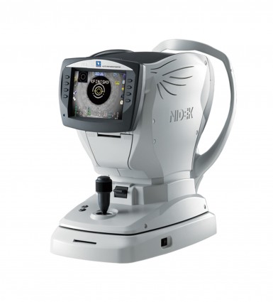
Auto Refractometer
An auto-refractometer is a computer-controlled machine used during an eye examination to provide an objective measurement of a person’s refractive error and prescription for glasses or contact lenses. This is achieved by measuring how light is changed as it enters a person’s eye. Auto-refractor calculates the refraction of the eye, sphere, cylinder and axis. This process is used to provide the starting point for the ophthalmologist or optometrist in subjective refraction tests. Automated refraction is particularly useful when dealing with non-communicative people such as young children or those with disabilities.
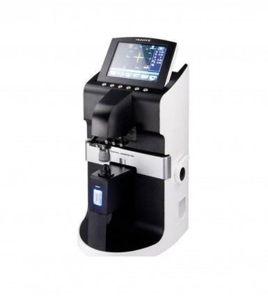
Auto Lensmeter
A lensmeter also known as a focimeter. It is an ophthalmic instrument, mainly used to verify the correct prescription in a pair of eyeglasses, to properly orient and mark uncut lenses, and to confirm the correct mounting of lenses in spectacle frames. Lensmeter can also verify the power of contact lenses, if a special lens support is used. The lensmeter is also used to check the accuracy of progressive lenses, and is often capable of marking the lens center and various other measurements critical to proper performance of the lens. It may also be used prior to an eye examination to obtain the last prescription the patient was given, in order to expedite the subsequent examination.
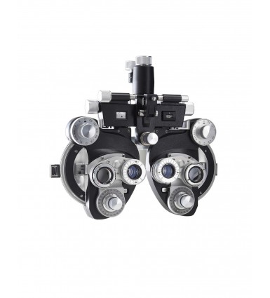
Manual Phoropter
A phoropter is an ophthalmic testing device. It is commonly used by eye care professionals during an eye examination, and contains different lenses used for refraction of the eye during sight testing, to measure an individual’s refractive error to determine the eyeglass prescription. Typically, the patient sits behind the phoropter, and looks through it at an eye chart placed at 6 metres, then near at around 40 centimetres (base on individuals preference reading distance) for reading glasses. The eye care professional then changes lenses and other settings, while asking the patient for subjective feedback on which settings gave the best vision.
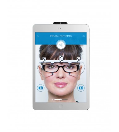
Essilor Eye Ruler 2
Eye Ruler 2 is an essential tool for dispensing personalized lenses. This digital ruler was designed and produced by Essilor International, a French-based international ophthalmic optics company that designs and manufactures ophthalmic lenses. Eye Ruler 2 is an accurate and compact optical measuring device that attach with an iPad. Eye Ruler 2 enable our optometrist to capture entirely new measurement as near vision behaviour, distance between two eyes, the positions of a frame on face, frame wrapped angle and etc. Its accuracy is up to 0.5mm and these measurements are essential for us to deliver best possible vision and best postural comfort personalized lenses as progressive lenses.
Corneal Pachymetry
A pachymetry is a simple, quick, painless test to measure the thickness of your cornea. With this measurement, our optometrist can better understand your Intraocular pressure (IOP) reading, and refer for a treatment plan that is right for your condition. The procedure takes only about a minute to measure both eyes. Corneal center thickness (CCT) is important because it can mask an accurate reading of eye pressure. Actual IOP may be underestimated in patients with thinner CCT, and overestimated in patients with thicker CCT. This is dangerous because if actual IOP is higher than reading shows, one may be at risk for developing glaucoma and doctor may not know it. Left untreated, high IOP can lead to glaucoma and vision loss.

I-care Handheld Tonometer
I-Care is a handheld contact tonometer. Tonometry is a diagnostic test that measures the pressure inside your eye, which is called intraocular pressure (IOP). There is no pain during the procedure. Tonometry is extremely safe. This measurement can help our optometrist to determine whether or not you may be at risk of glaucoma. Glaucoma is a serious eye disease in which there’s an increased fluid pressure within your eye. This increased pressure can damage your optic nerve. Since glaucoma can cause eventual blindness if it’s not treated, a tonometry test is critical for detecting eye changes early. If your test results come back abnormal, an early referral to eye doctor will be done to begin the treatment process, which can delay the progression of the disease.
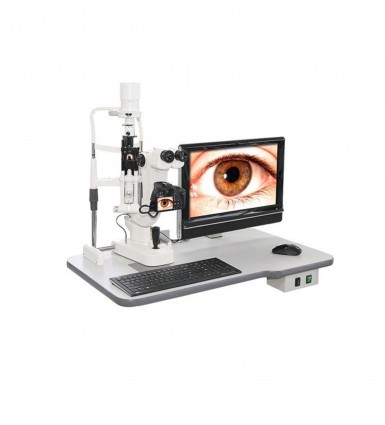
Slit Lamp Biomicroscope
Biomicroscope also known as slit lamp, is an instrument consisting of a high-intensity light source that can be focused to shine a thin sheet of light into the eye. It is used in conjunction with a biomicroscope. The lamp facilities an examination of the anterior segment and posterior segment of the eye, which includes the eyelid, sclera, conjunctiva, iris, crystalline lens and cornea. The binocular slit-lamp examination provides a stereoscopic magnified view of eye structures in details, enabling anatomical diagnoses to be made for a variety of eye conditions.

Corneal Topographer
Corneal topographer, also known as photo-keratometer. It is a non-invasive medical imaging technique for mapping the surface curvature of the cornea, the outer structure of the eye. Since the cornea is responsible for 70% of eyes refractive power, Its topograph is of critical importance in determining the quality of vision and cornea health. The three-dimensional map is a valuable in the diagnosis and treatment of a number of eye conditions and evaluating results and assessing the fit of contact lenses.

Optical Coherence Tomography
Optical coherence tomography (OCT) Is a non-invasive imaging test. OCT uses low-coherence light waves to capture micrometer resolution cross-section images of the retina, optic nerve head and anterior eye as cornea and anterior chamber angle. With OCT we can take a step further, our optometrist able to see each of the retina’s distinctive layers and this allows our optometrist to map and measure their thickness. These measurements help with diagnosis. They also provide treatment guidance for glaucoma and diseases of the retina.

Digital Fundus Camera
Fundus camera is a complex optical system used for imaging the retina of the eye. The main structures that can be visualized on a fundus photo are the central and peripheral retina, optic disc and macula. Fundus photos are ocular documentation that record the appearance of a patient’s retina. The photographs allow our optometrist to study a patient’s retina, detect retinal changes and review a patient’s retinal findings with a coworker. Fundus photographs are routinely called upon in a wide variety of ophthalmic conditions.
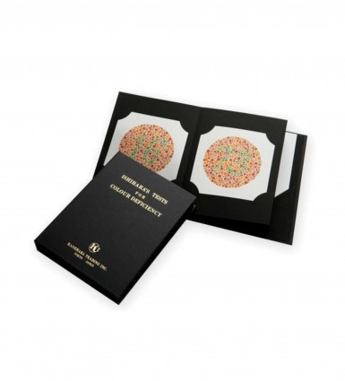
Ishihara 38 Plates Colour Deficiency Test
Ishihara test is a color perception test for red-green color deficiencies. The test consists of a number of colored plates, called Ishihara plates, each of which contains a circle of dots appearing randomized in color and size. Within the pattern are dots which form a number or shape clearly visible to those with normal color vision, and invisible, or difficult to see, to those with a red-green color vision defect. Other plates are intentionally designed to reveal numbers only to those with a red-green color vision deficiency, and be invisible to those with normal red-green color vision.
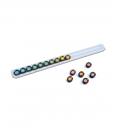
Farnsworth D15 Colour Vision Test
Farnsworth D15 are used to provide a detailed analysis of color blindness and/or a person’s ability to accurately perceive colors. These color blind tests consists of a certain number of colored discs or plates which have to be arranged in the correct color order. It contains 15 color discs, which need to be arranged in the correct color coded order. Patient must arrange the discs within the tray to create a continuum of gradually changing hue. Farnsworth D15 Test is useful for detecting protan, deutan and tritan colour defects. A disadvantage is, that minor color vision defects can not be detected.

Bellus 3D Face Scanner
Bellus 3D scanner scan of your face directly in our android app to capture over 20,000 millimeter accurate measurements of your face, including depth, width, and precise location of each ear. Allows us to create a precise model of your entire facial structure in the app, than you can select your style, choose your color, fine-tune the shape based on your personal preferences to make them distinctly you, and distinctly yours and preview it with our virtual try-on before we send for ordering. Your glasses are individually printed with 3D printer so that they never have to be heated, molded, or fitted to your face, and never lose their shape.
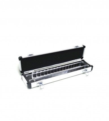
Progressive Lens Trial Set
We have a set of 10 pairs of progressive power lenses for purpose of trial, plastic lenses in metal shell rims from addition power of +1.00 D to +3.00 D in 0.50D steps to be used with adjustable PD trial frame to try it on as a progressive lens simulator. It provides patients the experience of vision with progressive lenses before order may help our optometrist to manage patient expectations better. This trial lenses ease the challenge in explaining multifocality to patients and would also ease their decision-making process. The progressive trial lenses provides practical utility in the our practice, in that it improves patient satisfaction, facilitates comparison of corrective solutions, and saves time by providing an easy explanation of multifocality.
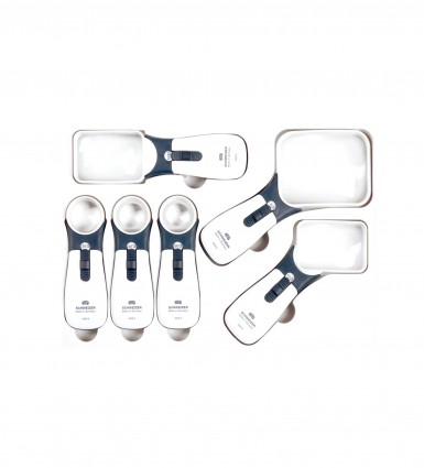
Handheld Magnifier
We carry a range of hand-held magnifier, including illuminated magnifier, stand-magnifier and pocket magnifier for vision rehabilitation. It is a process of restoring functional ability and improving quality of life and independence in an individual who has lost visual function through illness or injury. Most visual rehabilitation services are focused on low vision, which is a visual impairment that cannot be fully corrected by regular eyeglasses, contact lenses, medication, or surgery. This rehabilitation helps patients achieve physical, social, emotional, spiritual independence and quality of life. Rehabilitation does not undo or reverse the cause of damage; it seeks to promote function and independence through adaptation.
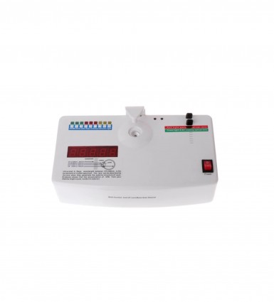
Ultra-Violet Tester
This ultraviolet (UV) tester work by detecting the UV radiation emitted through an ophthalmic lenses. To properly protect the eyes from the dangers of UV light, sunglasses should have UV-400 blocker to provide good coverage against the entire light spectrum that poses a danger. It is important to note that dark glasses that do not block UV radiation can be more damaging to the eyes than not wearing eye protection at all, because they tend to open the pupil and allow more UV rays into the eye. The UV protection of a lenses can be measured by this UV tester.
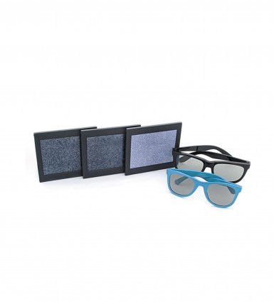
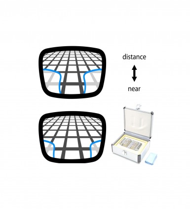
Random Dot "E" Stereopsis Test
The ability of stereopsis can be tested by this Random dot “E” stereogram. Random-dot stereopsis tests use pictures of stereo figures that are embedded in a background of random dots. Without stereopsis, the image looks only like a field of random dots. The random dot stereogram E (RDE) has been shown to be a simple and effective test for the detection of binocular abnormalities and defective visual acuity in children. The RDE is also reliable in screening primary school children to detect amblyopia and strabismus.

Retinoscope & Ophthalmoscope
Retinoscopy (Ret) is a technique to obtain an objective measurement of the refractive error of a patient’s eyes. The examiner uses a retinoscope to shine light into the patient’s eye and observes the reflection (reflex) off the patient’s retina. While, ophthalmoscopy is a test that allows a health professional to see inside the fundus of the eye and other structures using an ophthalmoscope. It is done as part of an eye examination and may be done as part of a routine physical examination. It is crucial in determining the health of the retina, optic disc, and vitreous humor.
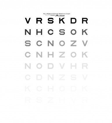
Pelli-Robson Contrast Sensitivity Test
Pelli-Robson chart consists of uniform-sized but increasingly pale gray letters on a white background, it is used to assess a patient’s contrast sensitivity. A contrast sensitivity test measures the ability to distinguish between finer and finer increments of light versus dark (contrast). This differs from common visual acuity testing in a routine eye exam, which measures your ability to recognize smaller and smaller letters on a standard eye chart. Contrast sensitivity is a very important measure of visual function, especially in situations of low light, fog or glare, when the contrast between objects and their background often is reduced. Driving at night is an example of an activity that requires good contrast sensitivity for safety.
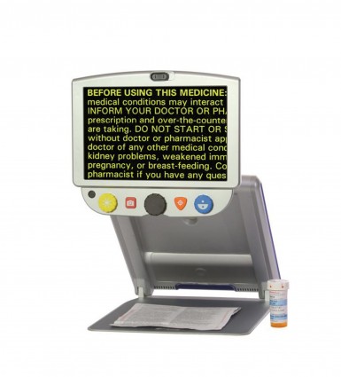
Portable Video Magnifier
Video magnifier is a reading aid for people with low vision that maximizes the remaining sight so it’s possible to read again. A video magnifier is an electronic magnifier that uses a camera and a screen to make text easier to read. The camera image will be magnified and the contrast can be increased, making it even easier to read. It also come with a portable size. A handheld video magnifier is a small, lightweight electronic magnifier that is easy-to- use. Due to its compact size, it is easy to carry along anywhere you go. You can magnify and increase contrast on the spot. Use a portable video magnifier on the go to read price labels, maps, bus schedules, directions and your favorite restaurant menu.
Horizontal & Vertical Prism Bars
These prism bar made from durable, lightweight acrylic. The horizontal prism bar provides 16 segments and the vertical prism bar has 15 segments The Combination Horizontal Vertical Prism Bar provides accurate measurement of ocular deviation. This horizontal and vertical prism bars is one of an instruments that required when performing a prism cover test. The prism cover test, is an objective measurement and the gold standard in measuring strabismus, i.e. ocular misalignment, or a deviation of the eye. It is used by optometrist and orthoptists in order to measure the vertical and horizontal deviation and includes both manifest and latent components.